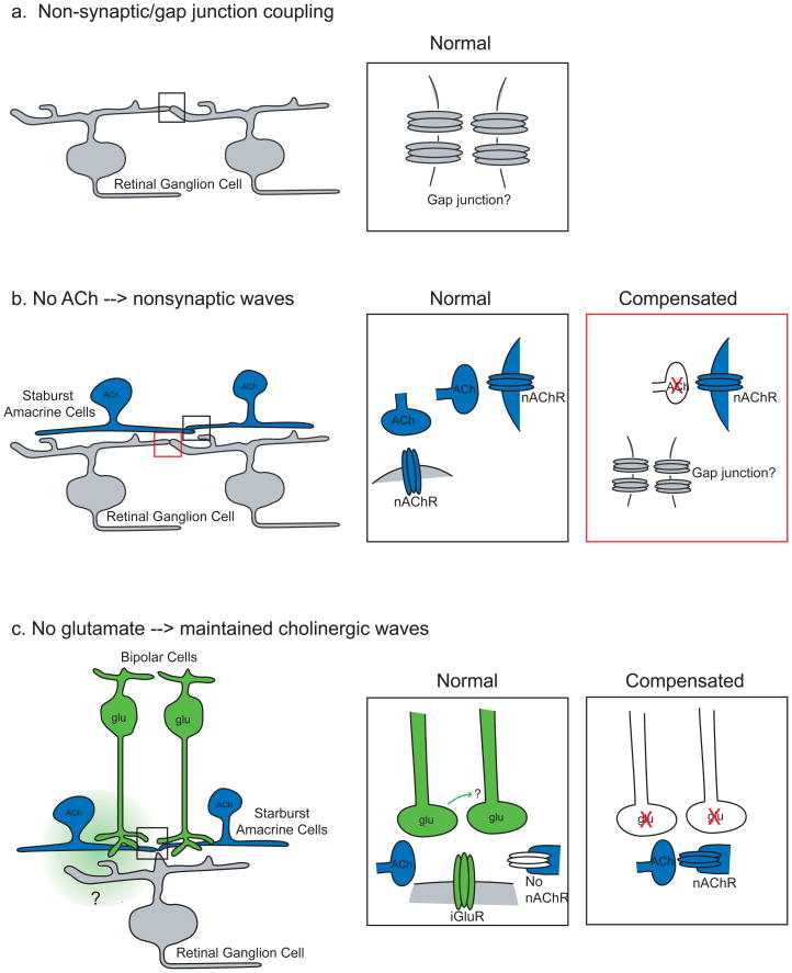
Figure 2. Homeostatic regulation of spontaneous network activity in the mammalian retina.
In the absence of a requisite circuit component, the retina regresses to the previous wave-generating mechanism. Here we provide schematics of the circuits that mediate retinal waves at different ages, including the changes that are thought to take place when one form of activity is disrupted.
a. Perinatally in mice, waves are mediated by a non-synaptic circuit, thought to be mediated by gap junction coupling (inset). Here the coupling is shown to be between retinal ganglion cells, although the location of the relevant coupling is not known.
b. During the first postnatal week, starburst amacrine cells (blue) form synaptic connections with other starburst amacrine cells and retinal ganglion cells (gray). Retinas from mice lacking acetyl choline (Ach; bottom inset) exhibit non-synaptic waves112, potentially through a reactivation of non-synaptic connections that mediate network activity in the perinatal period (see panel a). Furthermore, blocking nAChRs soon after the onset of cholinergic waves leads to the reappearance of non-synaptic waves97.
c: In the few days before eye opening in mice, when glutamatergic interneurons begin to form synapses with their postsynaptic targets, waves are mediated by glutamatergic circuits. Inset: Glutamatergic bipolar cells (green), which make glutamatergic synapses onto amacrine and ganglion cells and have no direct connections with each other, release glutamate that is detected both synaptically and extrasynaptically87. After the first postnatal week, starburst cells no longer express nAChRs56. Retinas from mice in which bipolar cells do not release glutamate (bottom inset) exhibit waves that are mediated by the cholinergic network87.

