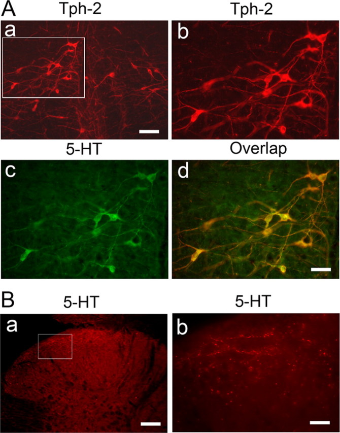
Figure 1.

Immunostaining shows the colocalization of Tph-2 with 5-HT in neurons of the RVM (A) and the distribution of 5-HT fibers in the spinal dorsal horn (B). Aa, Normal distribution of Tph-2-labeled neurons in low power of a tissue section at the rostral medulla level of the naive rats (n = 3). Ab is enlarged from the inset (Aa), illustrating the distribution of Tph-2-immunoreactive neurons in the RVM. Ac, 5-HT immunostaining in RVM neurons shown in Ab. Ad, Expression of Tph-2 with 5-HT in the same RVM neurons. Ba, 5-HT-labeled fibers and terminals in spinal dorsal horn of the naive rats (n = 3) at L5 level. Bb, An enlarged image of 5-HT fibers in the superficial dorsal horn cord from the insets (Ba). Scale bars: 100 μm (Aa, Ab, Ba), 50 μm (Ac, Ad), 20 μm (Bb).
