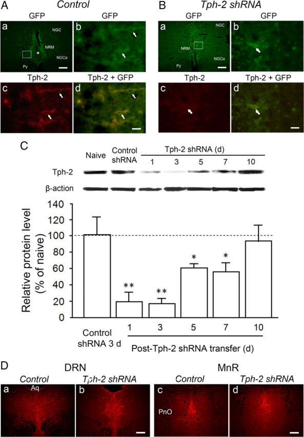
Figure 2.

Expression of GFP and Tph-2 immunostaining in RVM neurons after electroporation transfer of plasmids and RNAi with Tph-2 shRNA or control shRNA. A, GFP expression in the RVM neurons (arrows, Ab) at 3 d after control gene transfer is magnified from the inset in Aa. The asterisk (Aa) indicates the track of microinjection needle. Normal Tph-2 immunoreactivity (Ac) is present in GFP-expressing neurons (Ad). B, GFP expression in the RVM neurons (arrows, Bb) at 3 d after treatment with Tph-2 shRNA is enlarged from the inset in Ba. However, compared with the immunostaining in Ac, significant decrease or loss of Tph-2 immunoreactivity in the RVM neurons (Bc) is shown in the GFP-expressing cells (arrow, Bb–Bd). C, Western blot illustrating long-lasting decrease or depletion in Tph-2 in the RVM tissues from 1 d to 7 d after gene transfer [n = 3, *p < 0.05; **p < 0.01, vs control shRNA (n = 3)]. D, 5-HT immunostaining in the neighboring serotonergic nuclei, including the DRN (Da, Db) and the median raphe nucleus (MnR, Dc, Dd), at 4 d after control (Da, Dc) or Tph-2 shRNA (Db, Dd) treatment in the RVM. Scale bars: 100 μm (Aa, Ba, Da–Dd); 30 μm (Ab–Ad, Bb–Bd). Aq, Aqueduct; PnO, pontine reticular nucleus, oral part; Py, pyramidal tract.
