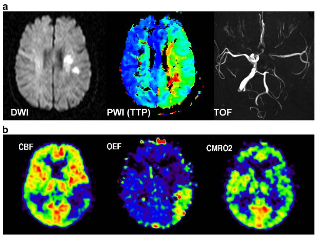
Fig. 1.
MRI and PET parameters used in the assessment of acute cerebral ischaemia. In a, diffusion-weighted imaging (DWI) depicts the area reflecting the core of infarction, perfusion-weighted imaging (PWI) reveals hypoperfusion in the territory of the middle cerebral artery (MCA), which is larger than the DWI lesion (mismatch), and time of flight (TOF) angiography shows a proximal occlusion of the MCA, which is perfused by leptomeningeal anastomoses. In b, cerebral blood flow (CBF) is measured by 15O-H2O PET, demonstrating hypoperfusion in the posterior portion of the MCA territory. By virtue of an increase in the oxygen extraction fraction (OEF), the cerebral metabolic rate of oxygen (CMRO2) as measured by 15O-O2 PET is still intact. This hypoperfused tissue, which is functionally disturbed but still viable, is the classic definition of penumbra [3]

