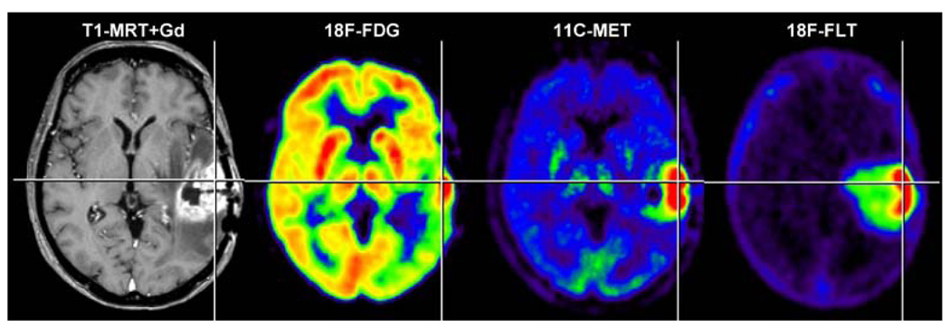
Fig. 4.
Parameters of interest in the non-invasive diagnosis of brain tumours. Alteration of the blood-brain barrier and the extent of peritumoural oedema are detected by MRI. Signs of increased cell proliferation can be observed by means of multitracer PET imaging using 18F-FDG, 11C-MET and 18F-FLT as specific tracers for glucose consumption, amino acid transport and DNA synthesis, respectively. Secondary phenomena, such as inactivation of ipsilat-eral cortical cerebral glucose metabolism, may be observed (18F-FDG) and are of prognostic relevance. Gd gadolinium. (With permission from [257])

