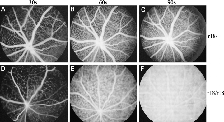
Figure 2.
Fluorescein angiograms of 3-week-old heterozygous and homozygous r18 mutant littermates. (A–C) The heterozygous mutant mouse shows normal retinal vasculature at 30 s (A), 60 s (B) and 90 s (C) after the injection of fluorescein dye. (D–F) The homozygous mutant mouse shows attenuated retinal vasculature and leakage of fluorescein dye in the retina detected at 60 s after the injection (E). The retinal vasculature becomes indistinguishable from the hyperfluorescent background at 90 s after the injection (F).

