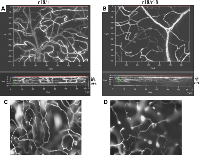
Figure 4.
Three-dimensional images of retinal vasculature labeled with fluorescent dye in 6-week-old r18 littermates. (A and B) A normal three-layer vessel network is present in the heterozygous control retina (A), whereas an attenuated and disorganized vessel network is present in the homozygous r18 mutant retina (B). Upper panels are front views of 3D retinal vasculature. The lower panels are rotated 90° relative to the upper panels. The bottom, middle and upper layers represent vessels in the OPL, IPL and GC layer, respectively. (C and D) Vessel networks are located in the OPL of heterozygous (C) and homozygous (D) retinas. Note the fully established vessel network in the heterozygous mutant retina, but attenuated vessels of incomplete network in the homozygous mutant retina.

