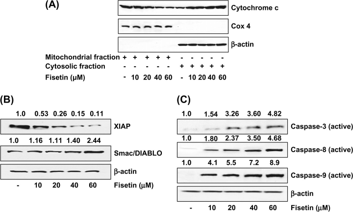
Fig. 5.
(A) Effect of fisetin on mitochondrial release of cytochrome c into cytosol. As detailed in Materials and methods, the cells were treated with fisetin (10–60 μM, 48 h) and then harvested. Mitochondrial and cytosolic fractions were prepared according to vendor's protocol and analyzed for cytochrome c and cytochrome c oxidase-4 by immunoblot analysis and chemiluminescence detection. The immunoblots shown here are representative of three independent experiments with similar results. (B) Effect of fisetin on protein expression of XIAP and Smac/DIABLO in LNCaP cells. (C) Effect of fisetin on protein expression of caspases-3, -8 and -9 in LNCaP cells. As detailed in Materials and methods, the cells were treated with fisetin (10–60 μM, 48 h) and then harvested. Total cell lysates were prepared and 40 μg protein was subjected to sodium dodecyl sulfate–polyacrylamide gel electrophoresis followed by immunoblot analysis and chemiluminescence detection. Equal loading of protein was confirmed by stripping the immunoblot and reprobing it for β-actin. The immunoblots shown here are representative of three independent experiments with similar results. The values above the figures represent relative density of the bands normalized to β-actin.

