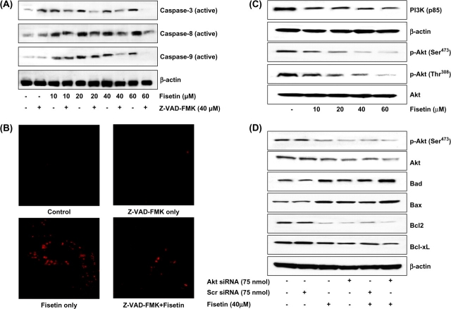
Fig. 6.
(A) Effect of Z-VAD-FMK on fisetin-induced activation of caspases in LNCaP cells. As detailed in Materials and methods, cells were incubated with 40 μM concentration of the general caspase inhibitor Z-VAD-FMK for 2 h followed by addition of fisetin (10–60 μM, 48 h) and then harvested. (B) Immunofluorescence staining for caspases-3 and -7. LNCaP cells were incubated with 40 μM concentration of the general caspase inhibitor Z-VAD-FMK for 2 h followed by addition of fisetin (40 μM, 48 h) and then harvested. Caspase activity was detected within whole living cells using ICT's Magic Red™ substrate-based MR-Caspase assay kit and fluorescence photographs were obtained at 510–560 nm excitation and 610 nm emission. The photomicrographs shown here are from one representative experiment repeated two times with similar results. (C). Inhibitory effects of fisetin on PI3K (p85) and phosphorylation of Akt (Ser473 and Thr308) in LNCaP cells. As detailed in Materials and methods, the cells were treated with fisetin (10–60 μM, 48 h) and then harvested. (D) Akt-dependent modulation in the Bcl-2 family proteins by fisetin in LNCaP cells. The LNCaP cells were transfected with Akt-siRNA (75 nM) or scrambled siRNA (75 nM) and were then treated with 40 μM fisetin for 48 h. Whole-cell lysate was prepared and 40 μg protein was subjected to sodium dodecyl sulfate–polyacrylamide gel electrophoresis followed by immunoblot analysis and chemiluminescence detection. Equal loading of protein was confirmed by stripping the immunoblot and reprobing it for β-actin. The immunoblots shown here are representative of three independent experiments with similar results.

