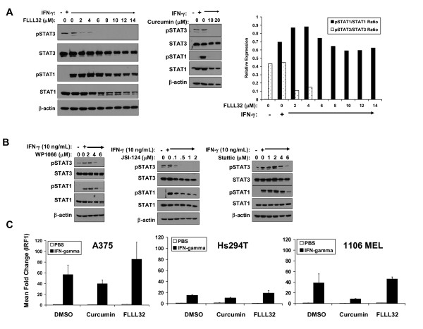
Figure 4.
IFN-γ-induced signal transduction was not adversely affected by FLLL32. (A) A375 cells were pre-treated for 16 hours with FLLL32 (2 -- 14 μM) or curcumin (20 μM) and subsequently treated with IFN-γ (10 ng/mL) for 15 minutes. IFN-γ-induced pSTAT1 and pSTAT3 were evaluated by immunoblot. Total STAT1, STAT3 and β-actin were also measured to control for loading. The data were also summarized by densitometry comparing relative expression of pSTAT1 to STAT1 and pSTAT3 to STAT3. (B) The same experiment was performed whereby A375 cells were pre-treated for 16 hours with other Jak2/STAT3 pathway inhibitors (WP1066, JSI-124, Stattic) prior to IFN-γ stimulation. Data shown are representative of two separate experiments and similar results were obtained in the Hs294T human melanoma cell line (Additional File 1: Figure S3). (C) IFN-γ-induced gene expression was enhanced in the presence of FLLL32. A375, Hs294T or 1106 MEL cells were pre-treated for 1 hour with 2μM FLLL32, 20μM curcumin or DMSO (negative control), and subsequently stimulated with IFN-γ (10 ng/mL) or PBS (vehicle) for an additional 4 hours. Expression of IRF1 was evaluated by Real Time PCR. Data were normalized to 18s rRNA levels (housekeeping gene) and expressed as the mean fold change versus DMSO-pre-treated cells stimulated with PBS. Error bars represent the standard deviation from n = 2 independent experiments.

