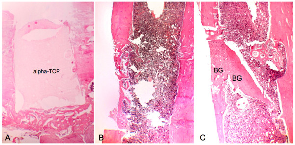
Figure 7.
Histological aspects of samples collected on postoperative day 21. A, T cavity, The alpha-tricalcium phosphate (alpha-TCP) block occupies all the bone cavity. The regular margins of the cavity contrast with the irregular surface of the material. (Magnification 40×). B, C - cavity, Continued healing of the cortex, in the upper surface of the surgical cavity. The medullary channel shows progressively increasing regularization. (Magnification 40×). C, C + cavity, The bone graft (BG) is seen in continuity with the trabecular area, which is merging into cortical bone. (Magnification 40×).

