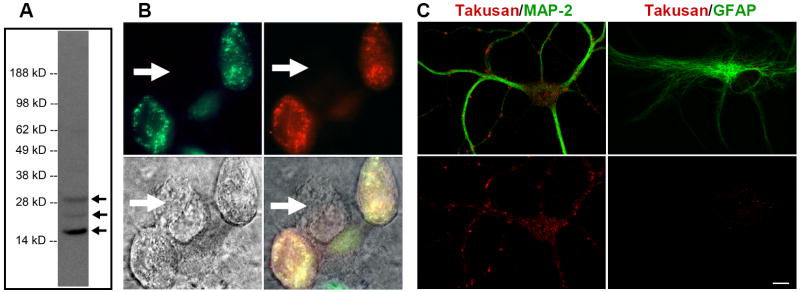
Figure 3. Characterization of Endogenous α-Takusan Proteins.
(A) Immunoblot of the PSD fraction prepared from adult C57BL/6 WT mouse brain using α-takusan antibody. The antibody detected a major band at ~17 kD and minor bands at ~24 and ~30 kD in this preparation. (B) Immunocytochemistry with α-takusan antibody of HEK293 cells expressing exogenous EGFP-α1. Images represent EGFP-α1 fluorescence (green, top left), anti-α-takusan staining (red, top right), Nomarsky (bottom left), and overlay (bottom right). Staining pattern of α-takusan approximates that of EGFP; additionally, a cell not expressing EGFP-α1 does not stain for α-takusan (indicated by the arrow). (C) Selective expression of α-takusan in MAP-2 positive neurons. Mixed neuronal/glial cultures of hippocampal cells were co-stained for α-takusan/MAP-2 or α-takusan/GFAP. The two images at the top represent an overlay of α-takusan (red) and either MAP-2 (green; left) or GFAP (green; right). The images on the bottom represent α-takusan staining only. The α-takusan antibody detected strong signals in MAP-2 positive (neuronal) cells, but only weak signals in GFAP-positive (astrocytic) cells. In the MAP-2 positive cells, α-takusan localized to both dendrites and cell bodies. Scale bar = 10 μm.

