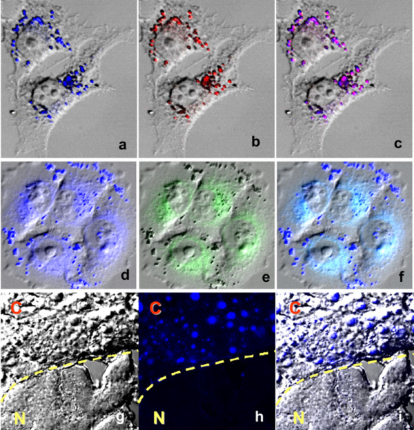
Figure 2.

Intracellular localization of AC-202. HT168 human melanoma cells were double-stained with the blue fluorescent AC-202 (a. 5 μM; d. 20 μM), lipid droplet-specific dye (oil red O) (b), and endoplasmic reticulum-specific dye (ER-Tracker™ Green) (e). Purple color derives from co-localization of oil red O and AC-202 (c). Cyan color derives from co-localization of ER-Tracker™ and AC-202 (f). Pictures d-f were recorded with higher laser power. In vivo staining of lipid droplets in prenecrotic cancer tissues (HT168 xenograft) in liver of SCID mouse by AC-202 (g-i). "C" denotes for cancer tissue, "N" for normal. Cancer and normal cells are separated by a dashed line.
