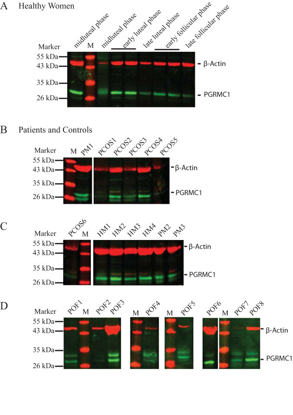
Figure 1.
Detection of PGRMC1 and β-actin by Western blot analysis in total protein preparations obtained from peripheral nucleated blood cells (PNBC). Representative western blot pictures illustrating detection in (A) healthy cycling women (HF; distinct phases are indicated above), (B-D) PCOS and POF patients and control groups (PM, natural menopausal women; HM, healthy men). Total protein preparations were separated on a 12% SDS-PAGE, transferred to a PVDF membrane and subsequently detected using α-PGRMC1 and α-β-actin antibodies. A protein standard was included and the band sizes are indicated to the left. Bands corresponding to PGRMC1 (green) and β-actin (red) are indicated.

