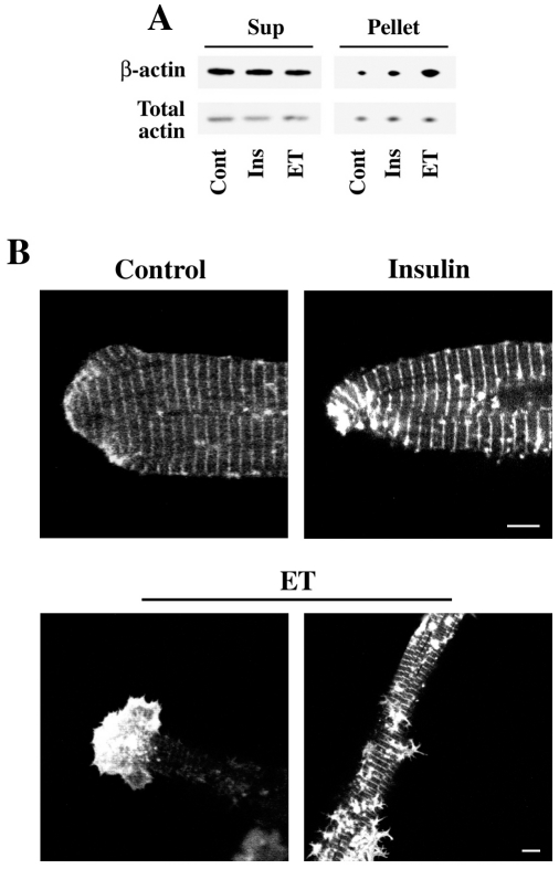
Figure 3. β-actin cytoskeletal reorganization during hypertrophic stimulation in cultured cardiomyocytes.
A) Adult cardiomyocytes cultured on laminin-coated dishes were treated with 200 nM endothelin (ET) or 100 nM insulin (Ins) for 30 min. The cells were lysed and subjected to F-actin (Pellet) and G-actin (Sup) fractionation as described under Materials and Methods. Both these fractions were analyzed by Western blotting using indicated antibodies. B) Adult cardiomyocytes infected with β-actin-GFP were treated with insulin (100 nM) or endothelin (200 nM) for 48 h and imaged at various Z-planes using a confocal microscope. Scale bar = 5 µm.

