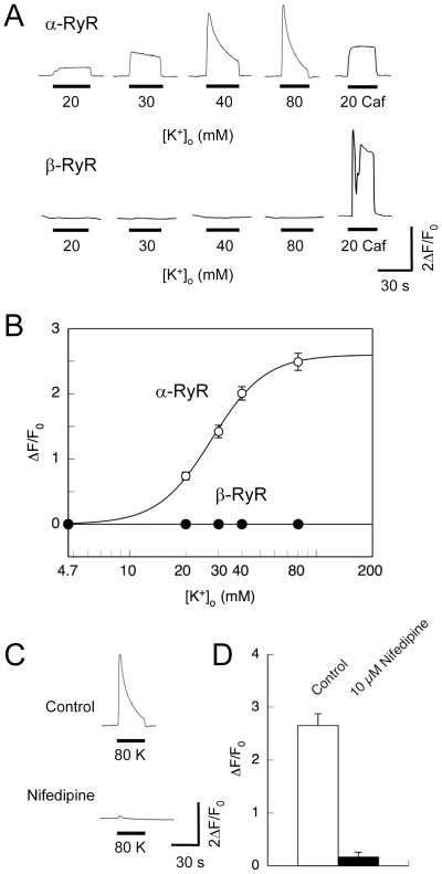
Figure 2. Depolarization-induced Ca2+ release.
Intracellular Ca2+ ([Ca2+]i) of 1B5 myotubes transduced with either α-RyR or β-RyR virions was imaged using fluo-4 as described in Materials and Methods. Cells were exposed for 30 sec to varied high [K+]o solutions (with the constant [K+]×[Cl−] product) to trigger DICR, and finally to 20 mM caffeine to confirm the functional expression of RyR. Ca2+ in the bath solution was omitted prior to and during stimuli to prevent Ca2+ influx. A. Representative traces of Ca2+ transients of individual cells by [K+]o and caffeine. B. Averaged maximal change in fluo-4 fluorescence (ΔF/F0) was plotted against [K+]o concentration. Values are expressed as mean ± SE (n = 112 for α-RyR and 18 for β-RyR). α-RyR but not β-RyR exhibited Ca2+ transients induced by high [K+]o. C. A representative trace of Ca2+ transients of myotubes expressing α-RyR stimulated with 80 mM [K+]o before and after treatment with 10 µM nifedipine. D. Averaged maximal change in fluo-4 fluorescence (ΔF/F0) was plotted with or without 10 µM nifedipine. Values are expressed as mean ± SE (n = 15). Nifedipine inhibited high [K+]o-induced Ca2+ transients.

