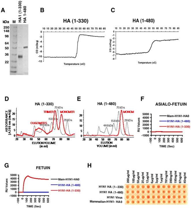
Figure 1. Biochemical and functional characterization of bacterially expressed and purified H1N1 HA proteins.
(A) Purified E. coli derived HA proteins were analyzed by SDS-PAGE. DNA encoding HA1 (1–330) and HA (1–480) from HA gene segment of A/California/07/2009 (H1N1) generated from egg-grown virus were used for cloning in a T7 promoter based expression vector (pSK) where the desired polypeptide can be expressed as fusion protein with His6 tag at the C-terminus. The proteins were expressed, denatured and refolded under controlled redox conditions and purified using His-Trap fast flow chromatography to >90% purity (see Materials and Methods). The purified proteins run at their corresponding molecular weight in reducing SDS-PAGE. (B-C) CD melt spectroscopy shows that both H1N1 HA1 (1–330) (B) and H1N1 HA (1–480) (C) are properly folded. Both H1N1 HA proteins, at a concentration of 0.5 mg/ml in 20 mM PBS, pH 7.2, were subjected to heating at 0.5°C/min increments. The protein unfolding kinetics was measured at 222 nm using a J-715 Circular Dichroism system (JASCO corp., Easton, MD). (D-E) Superdex S-200 gel filtration chromatography of purified H1N1 HA proteins from E.coli. The panels present superimposed elution profiles of purified HA proteins (red line) overlaid with calibration standards (grey line). (D) The H1N1 HA (1–330) protein purified from bacterial cells existed as approximately 20% high-molecular-mass oligomer (>600 kDa), 45% trimer (∼110 kDa) and 35% monomer (34kDa) (red line). (E) H1N1 HA (1–480) is present only as a monomer (50kDa). (F-G) Binding kinetics of purified H1N1 HA proteins in a SPR based receptor binding assay. Steady-state equilibrium analysis of different H1N1-HA proteins to fetuin and its asialylated counterpart (Asialo-fetuin) was analyzed at 25°C using a ProteOn surface plasmon resonance biosensor (BioRad Labs). Samples of purified H1N1-HA proteins (10 µg/ml) were injected simultaneously over a mock surface to which no protein was bound, followed by the Asialofetuin in (F) or Fetuin in (G) immobilized on a sensor chip through the free amine group, and onto a blank flow cell, free of protein. Binding kinetics and data analysis were performed using ProteOn system surface plasmon resonance biosensor instrument (BioRad Labs, Hercules, CA). (H) Agglutination of human RBCs by properly folded bacterial H1N1 HA (1–330) protein. Serial dilutions of purified HA proteins or virus were mixed with washed RBC and incubated to analyze the receptor binding and cross-linking of human RBC. Virus H1N1xPR8 A/California/07/2009 (X-179A) was used as a control. Strong hemagglutination was observed for bacterial H1N1 HA (1–330) but not with either bacterial H1N1 HA (1–480) or mammalian H1N1 HA0.

