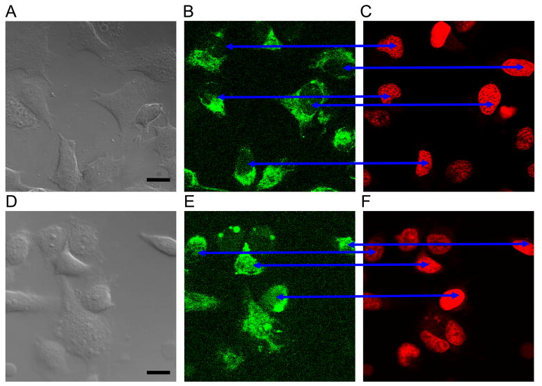
Fig 2.
Agent with AGUGUU penetrated some HeLa nuclei while agent without did not. Images A,D are DIC micrographs with bar = 10 um; images B,E are FAM emission confocal micrographs; images C,F are DRAQ5™ emission confocal micrographs. Cells in the upper three images were transfected with FAM+UAGCACCAUUUGAAAUC; no nuclei have FAM fluorescence. Cells in the lower three images were transfected with FAM+UAGCACCAUUUGAAAUCAGAGUU; most nuclei have at least some FAM fluorescence (blue arrows). Here and also in Figs 3 and 5 the brightness and contrast settings for each fluorescence channel are identical. The confocal images are from a ~400 nm slice of the sample. Cells were alive. The images were obtained 3 to 4 hours post-treatment, hence nuclear import was not always related to a particular cell cycle phase, including the mitosis phase. Scale bar = 10 μm.

