
Figure 2. Sox2 inactivation impairs proliferation of primary osteoblasts.
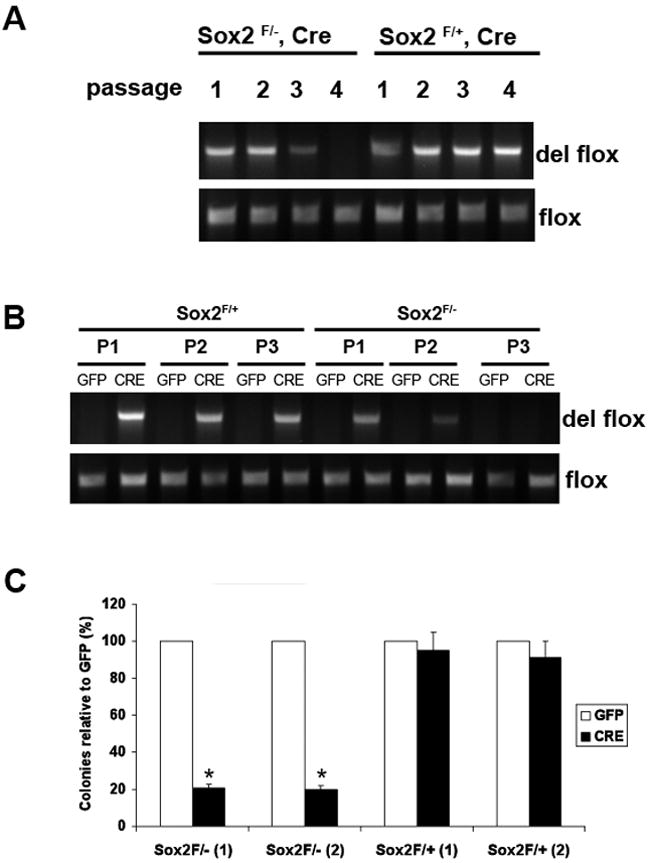
(A) PCR analysis of primary osteoblast DNA. Primary calvarial osteoblasts were prepared from Sox2flox/-;Cre (cko) mice and Sox2flox/+;Cre (control). Upon confluency, cells were passaged and DNA was extracted at each passage. The deleted flox and the flox allele were detected by PCR using specific primers to assess persistence of Sox2-null cells. Numbers indicate passage number following isolation.
(B) PCR analysis of GFP- and Cre-virus infected primary osteoblast DNA. Primary osteoblasts from Sox2 flox/+ (control) and Sox2 flox/- (cko) mice were infected with GFP or Cre-adenovirus and passaged after infection. DNA was extracted from each passage and the deleted flox allele was detected by PCR using specific primers to check for persistence of Sox2-null cells.
(C) Colony Assay. Primary calvarial osteoblasts from Sox2 flox/+ (control) and Sox2flox/- (cko) mice were infected with either GFP or Cre-adenovirus for 72 hours and plated in triplicate in 6-well plates. Colonies were counted after crystal violet staining. Percent of colonies obtained in Cre infection are plotted as a percentage of the colonies in the corresponding GFP infection in two independent Sox2 flox/- and Sox2 flox/+ osteoblast pools [(1) and (2)]. * = p<0.05. GFP Adenovirus infection of Sox2flox/- or Sox2flox/+ cells produced equivalent number of colonies (~120 colonies/plate).