Figure 1.
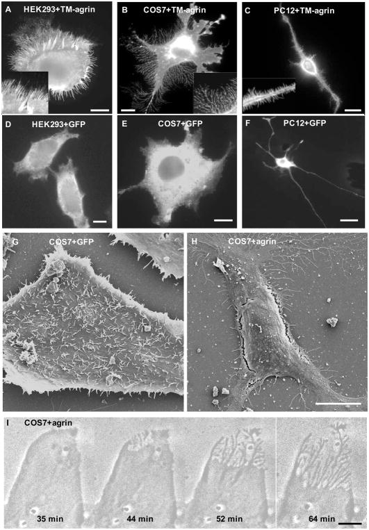
Over-expression of TM-agrin induces filopodia-like processes in HEK293, COS7 and PC12 cells. Cells were fixed 24 h after transfection with TM-agrin-GFP (A, B and C) or GFP (D, E and F). TM-agrin-GFP was detected by immunofluorescence staining with anti-GFP. All three cell lines showed extensive formation of filopodia-like processes (A–C) compared to control cells transfected with the GFP vector (D–F) but the morphology of the induced processes varied. Insets in A-C show the filopodia-like processes at higher magnification. (G, H) Scanning electron micrographs of COS7 cells fixed 24 h after transfection with GFP (G) or TM-agrin GFP (H). Many of the GFP-transfected cells had many filopodia on the unattached surface, whereas the TM-agrin-GFP transfected cells displayed BRFs and filopodia attached to the substratum but few if any filopodia on the unattached surface. (I) Phase contrast images from a time-lapse sequence of a COS7 cell starting 18 h after transfection with TM-agrin-GFP, showing that BRFs form by retraction of lamellipodia. All reference bars = 10 μm.

