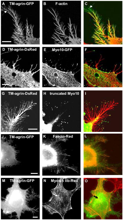
Figure 2.
BRFs induced by over-expression of TM-agrin-GFP in COS7 cells resemble filopodia in protein localization. (A) TM-agrin-GFP visualized by immunofluorescence staining of GFP (green). (B) F-actin labeled with rhodamine-phalloidin (red) is found throughout the BRFs. The distal GFP-positive areas lacking F-actin staining (arrows) represent TM-agrin-GFP shed onto the substratum before the tips of BRFs retracted. (D, G) TM-agrin-Ds-Red (red) visualized by immunofluorescence staining of Ds-Red. (E, H) Over-expressed myo10-GFP and truncated myo10-GFP, visualized by immunofluorescence staining with anti-GFP (green) were localized to the tips of BRFs. (J, M) Over-expressed TM-agrin visualized by immunofluorescence staining of GFP (green), (K) Fascin labeled with fascin antibody (red) is found in BRFs, (N) Myosin II-B labeled with myosin II-B antibody (red) is absent from BRFs, but present in stress fibers (arrows). (C, F, I, L, O) Overlay images. Reference bar = 10 μm.

