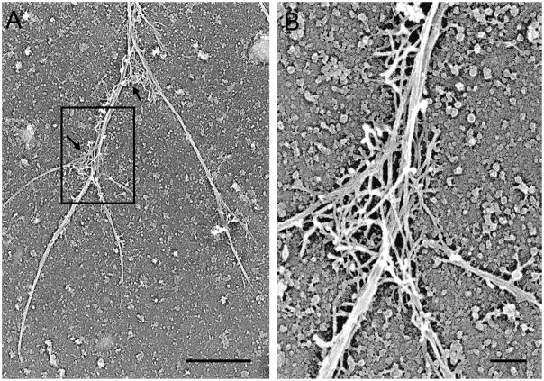
Figure 3.
BRF cytoskeletons consist of long actin filament cables, with actin filament networks primarily at branch points. Platinum replica electron micrograph of a detergent-extracted cytoskeleton shows a segment of a BRF cytoskeleton. Arrows in (A) indicate two branch points with actin filament networks resembling those found in lamellipodia. The marked area in (A) is shown at higher magnification in (B). Reference bars = 1μm and 0.2μm in (A) and (B), respectively.

