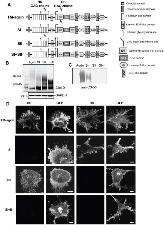
Figure 5.
Mutation of the GAG-chain attachment sites results in elimination of the expected GAG chain types in over-expressed TM-agrin. (A) Domain structures of TM-agrin-GFP and the GAG chain mutants used in this study. GFP was attached to the C-terminal end of these constructs. (B) The high molecular weight smear of glycosylated agrin is reduced both in range of molecular weight and in density relative to the agrin core protein in GAG chain mutants. COS7 cells overexpressing TM-agrin-GFP or the mutants were lysed 24 h after transfection and the proteins were separated by SDS-PAGE. The blots were stained with GFP antibody. A separate blot for GAPDH shows that protein loading was equivalent in all lanes, however, total agrin-GFP expression levels in this experiment varied, as indicated by variation in total GFP immunoreactivity. The agrin-GFP core protein band ran at ~ 220 kD. The narrower, lower molecular weight smear in the SI+II mutant is presumed to represent agrin with non-GAG glycosylation. (C) Chondroitin sulfate GAG chains are absent in SII and SI+II mutants. Western blots of wild-type and mutant lysates were stained with chondroitin sulfate antibody CS-56. (D) HS and CS GAGs are lacking in COS7 cells over-expressing the respective GAG chain mutants. Cells transfected with TM-agrin-GFP or mutants (SI, SII and SI+II) were fixed after 24 h and immunostained with mouse monoclonal HS (left-hand column) or CS (3rd column from left) antibodies followed by Cy3-conjugated anti-mouse. TM-agrin and mutants were detected by GFP fluorescence (2nd and 4th column from left). Cells were imaged by confocal microscopy. Cells over-expressing wild-type TM-agrin-GFP showed strong staining for HS and CS but the SI, SII and SI+II mutants lacked immunoreactivity with HS, CS and both antibodies, respectively. Note that GFP fluorescence indicated similar levels of membrane expression for the wild-type and mutant forms of TM-agrin-GFP. Reference bar = 10 μm.

