
Figure 2.
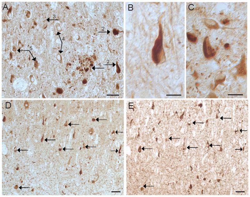
RTN3 in AD. In the hippocampus of a 67 year old AD case (A), RTN3 localized to dystrophic neurites around senile plaques (1), NFTs (2), neuropil threads (3), neuronal cytoplasm (4), and GVD (5). NFT are prominent in the majority of AD cases, including a 72 year old patient (B) and a 59 year old patient (C). On adjacent serial sections from an 80 year old case of AD stained for RTN3 (C) and AT8 (D), many of the same NFT contain both proteins (arrows). Scale bars= 50 μm (A,D,E), 20 μm (B,C,).