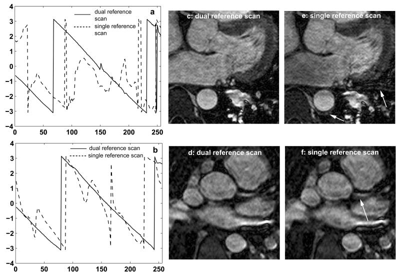
Figure 3.
Comparison between the dual and single reference scan phase correction techniques in 2 healthy volunteers (a, c, e: volunteer 1 and b, d, f: volunteer 2). a, b: Phase difference between the even and odd echoes. With the dual reference scan technique (solid line) the phase difference is linear, whereas with the single reference scan technique (dotted line) it deviates from the expected linear approximation. c-f: effect of dual (c, d) and single (e, f) reference scan phase correction techniques on the images. Ghosting and blurring artifacts shown by the white arrows in the images obtained with the single reference scan technique are eliminated by using the dual reference scan technique.

