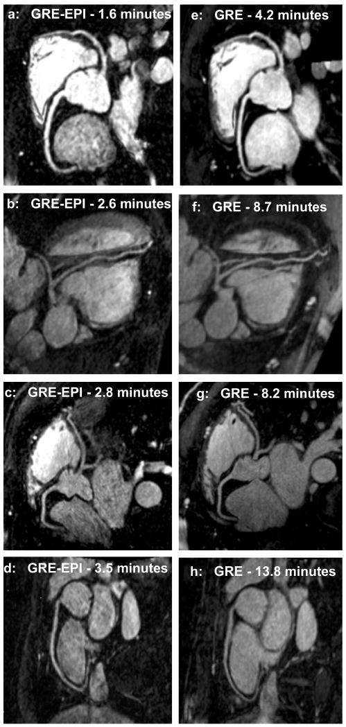
Figure 4.
Reformatted coronary artery images from 4 volunteers using a contrast-enhanced GRE-EPI sequence (4a-d) and a contrast-enhanced GRE sequence (4e-h), acquired in separate scan sessions. The GRE-EPI sequence has contrast agent dose reduced by a factor of 2 and scan time reduced by a factor of 3.

