
Figure 2. Structure and function of dendritic HCN channels.
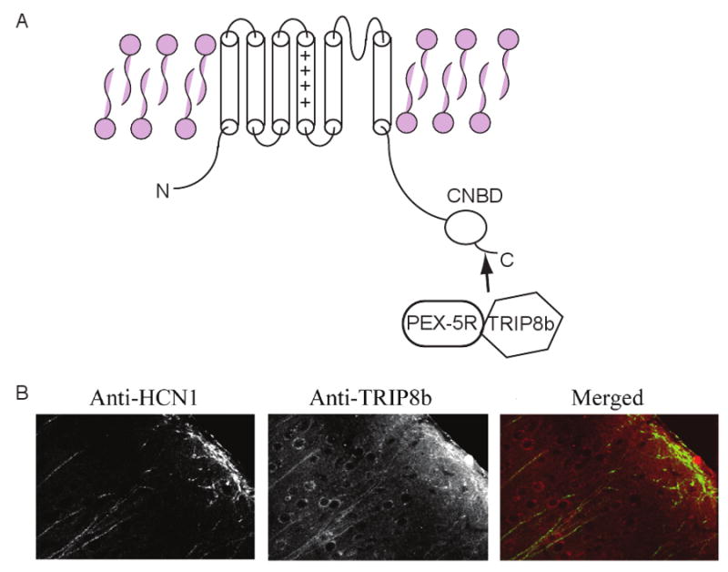
A) Illustration of TRIP8b and PEX5Rp interaction with the C-terminus of HCN subunits. The Cyclic Nucleotide Binding Domain (CNBD) which binds to cAMP is also located on the C-terminus. B) Confocal images showing TRIP8b and HCN1 expression in cortical layer V cell dendrites. The right panel shows the merged image of rhodamine-red labelled anti-TRIP8b and FITC labelled anti-HCN1. Co-localization is depicted as yellow on this image. The double staining was obtained in the same brain section (adapted from Ref. [92]).