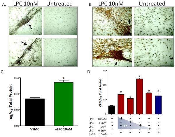
Figure 2. LPC-treated VSMCs produce calcium phosphate deposits in culture.
(A) von Kossa stain, 14d, 4X. Arrows indicate calcium phosphate ridges. (B) Alizarin Red S calcium stain, 14d, 4X. Arrows indicate calcium structures. Scale bar = 200μm (C) Total phosphate levels; n=3, p<0.05, phosphate (μg) normalized to total protein (μg) (D) Calcium incorporation assay; 45CaCl2, n=6,*p<0.01, #p<0.05. CPM was normalized to total protein (μg/μL) concentrations (CPM/μg total protein). Error bars represent ±SD.

