
Figure 2. Effects of tgfα pRNAi during oogenesis in T. castaneum, N. vitripennis, and G. bimaculatus.
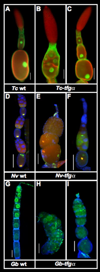
A–C: T. castaneum ovarioles stained with phalloidin (red) and α-nuclear pore antibody (green). In wild type, each oocyte becomes encapsulated by a single layer of follicle cells, and the oocyte nucleus moves to a cortical location about midway along the long axis of the oocyte in late oogenesis (A). In strongly affected Tc-tgfα pRNAi ovarioles, oocytes are not encapsulated, and form a mass of germline cells that are not separated by follicle cells. (B) In presumably weaker cases, and the position of the oocyte nucleus is disrupted by Tc-tgfα pRNAi (C). See also Fig S2.
D–F: Expression of DAPI (blue), Nv-otd1 (green) and Nv-nos (red) in wild type (D) and Nv-tgfα pRNAi (E,F) ovarioles from N. vitripennis. Nv-tgfα pRNAi ovarioles show defects in encapsulation of the oocytes by follicle cells (E), as well as improper localization of normally anteriorly and posteriorly localized mRNAs (E,F). In general there is also a major reduction in production of germline cells.
G–I: Ovarioles of G. bimaculatus stained for α-nuclear pore (green) and DAPI (blue). Egg chambers in wild type ovaraioles show a linear arrangement, and an increasingly elongated cylindrical shape during early oogenesis (G). In strongly affected Gb-tgfα pRNAi ovarioles (H), oocytes are arranged chaotically, and are not encapsulated by a complete layer of follicle cells. In moderately affected ovarioles, oocytes are encapsulated, but take on a more rounded shape (I).
Scale bars represent 100μm.