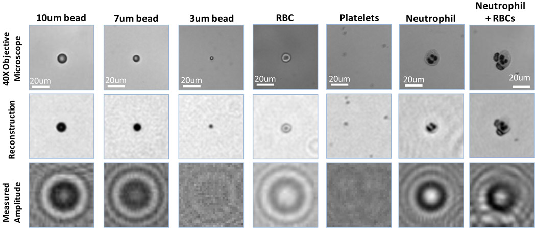
Fig. 3.
Various objects imaged using the lensfree incoherent holographic microscope of Fig. 1(a) are illustrated and compared against 40× objective-lens (NA=0.6) images of the same FOV. The bottom row illustrates the lensfree incoherent holograms that are digitally processed to reconstruct the middle row images. The last 3 columns are taken from a blood smear sample, whereas the other 4 columns on the left are imaged within a solution/buffer. Same imaging parameters as in Fig. 2 are used.

