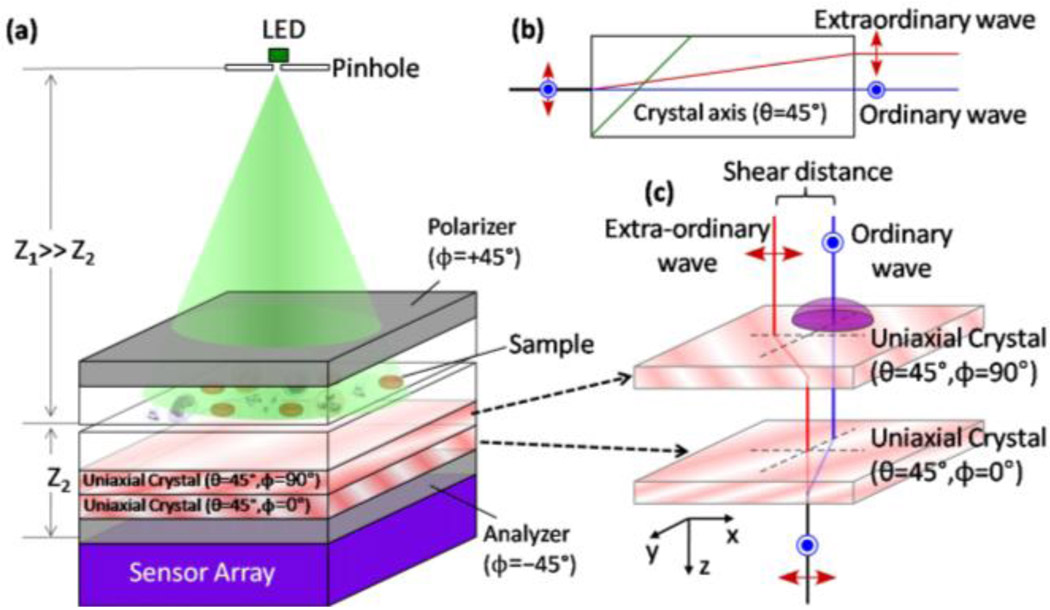
Fig. 4.
(a) Schematic diagram of the lensfree differential interference contrast (DIC) microscopy configuration is illustrated. The same holographic microscope of Fig. 1(a) can be converted into a lensless DIC microscope with two polarizers and two thin birefringent crystals as illustrated in (a). (b) Each birefringent uniaxial crystal creates an ordinary and an extra-ordinary wave of the object, which are separated by ~1.1 µm from each other for a quartz thickness of 0.18mm. This double refraction process is wavelength dependent, and to ensure zero net phase bias between the ordinary and the extra-ordinary holograms regardless of the LED wavelength, two crystals are glued to each other with 90° rotation in between as illustrated in (a) and (c). This sandwiched crystal assembly increases the total shear distance between the ordinary and the extra-ordinary holograms by a factor of √2, and together with the analyzer at 45°, it creates the DIC hologram of each object that is sampled by the sensor array. Despite the major differences in the way that the lensless holograms are created and recorded for regular transmission imaging vs DIC imaging, the digital image reconstruction process remains the same in both of the approaches.

