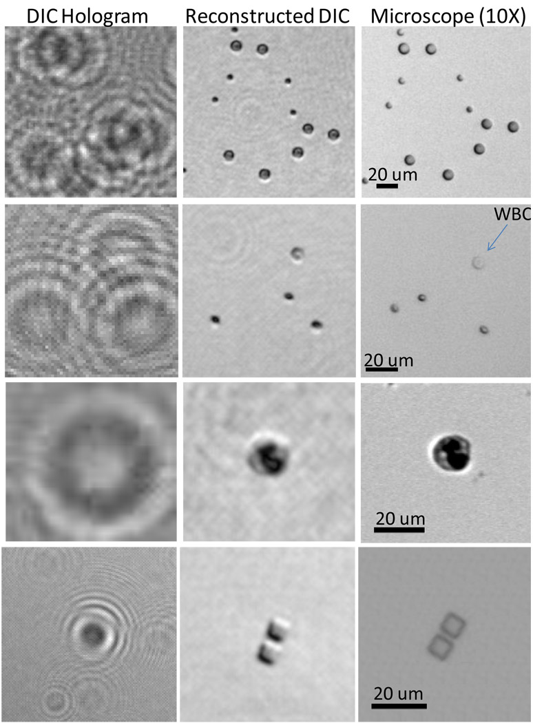
Fig. 5.
Differential interference contrast (DIC) images of various objects that are acquired with the handheld lensless holographic microscope of Fig. 1(a) are illustrated. Top Row: 5 and 10 µm polystyrene particles; 2nd Row: white and red blood cells in diluted whole blood (aqueous); 3rd Row: A white blood cell on a blood smear; 4th Row: FIB etched square structures that are separated by 2 µm on a glass substrate. For these images, 2 plastic polarizers and 2 thin quartz crystals were added to the lensless microscope of Fig. 1(a), as illustrated in Fig. 4. The total cost of these additional components is < 2 USD. D=100 µm, z1=~3.5 cm, z2=~1.3 mm and an integration time of ~600 ms were used for capture of the raw DIC holograms. Relatively increased integration time in these DIC images is due to the cross-polarizer configuration as shown in Fig. 4(a). 10× objective-lens (NA=0.2) microscope images of the same objects are also illustrated at the right column. For the relative positions of the micro-particles/cells in a solution, there are some unavoidable shifts between the lensless DIC image and the microscope comparison due to movement of the particles/cells between the capture of each comparison image.

