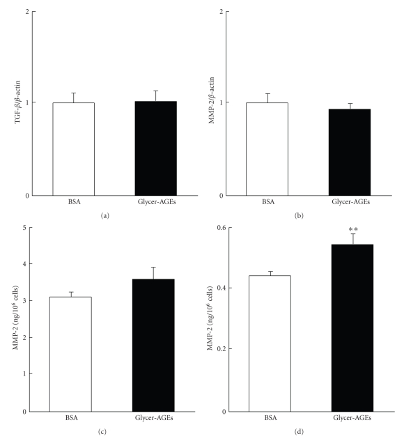
Figure 5.
(a) and (b) Cells were incubated with control unglycated BSA or Glycer-AGEs for 24 h. The levels of TGF-β (a) and MMP-2 (b) mRNA expression were analyzed using the real-time RT-PCR method, and the result was normalized to the β-actin mRNA level. (c) and (d) Cells were incubated with control unglycated BSA or Glycer-AGEs for 48 h. The conditioned medium was collected, and the activities of pro-MMP-2 (c) and MMP-2 (the activated form) were measured (d). Data are shown as the mean ± SD (n = 3) **P < .01 versus control unglycated BSA.

