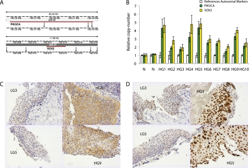
Figure 3.
(A) Multiple markers were designed to interrogate the copy number of two genes, PIK3CA (n = 3) and SOX2 (n = 5); the markers' location is indicated by black arrows in a modified screenshot from the Ensembl database. (B) The copy number of PIK3CA and SOX2 was analyzed for each high-grade dysplasia (HGD) and pooled normal genomic DNA (N). Results from individual experiments show the mean and standard deviation of multiple markers for each locus (reference autosomal, [n = 3–5]; PIK3CA and SOX2). (C) Immunohistochemistry for PI3Kα on low-grade dysplasia (LGD) and HGD biopsies. In seven of nine biopsies there was strong cytoplasmic staining in HGD compared with weak staining in five of seven LGD lesions. (D) Nuclear expression of SOX2 was increased in a number of high-grade lesions relative to low-grade lesions. However, for a number of lesions it was not possible to discriminate LGD and HGD on the basis of SOX2 immunostaining.

