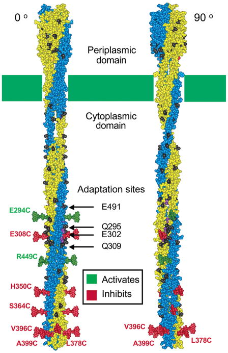
Figure 4.
Two orientations of the receptor structural model (2, 8) illustrating the eight positions at which 5-fluorescein-maleimide (5FM) probe attachment yields the largest perturbations of receptor-stimulated kinase activity. The two orientations differ by a 90° rotation about the long axis of the dimer. Shown at each perturbing position is a plausible orientation for a 5FM probe coupled to that position. Green and red probes indicate positions at which 5FM attachment strongly superactivates or inhibits the kinase, respectively. Prominent in the left orientation are the two symmetric linear interaction faces that contain the adaptation sites, while in the right orientation the two symmetric radial interaction faces are more prominent.

