Figure 6.

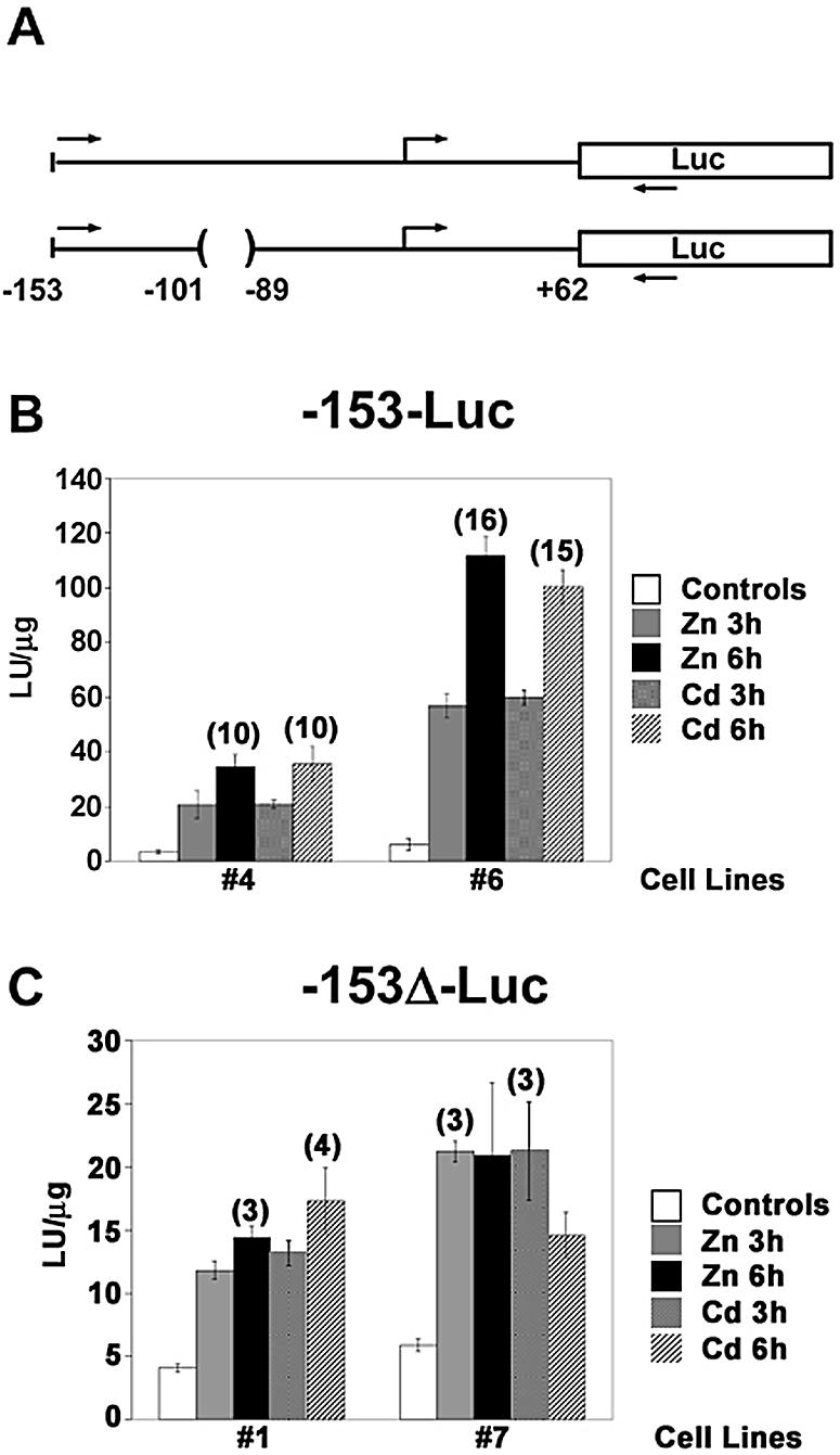
Characterization of stably-transfected cells used for in vivo mapping studies. (A) Diagram of the MT-I promoter-Luciferase fusion vectors. Structures of the Luciferase (Luc) reporter genes under the control of a minimal (–153 to +62 bp) MT-I promoter with (top) or without (bottom) the USF/ARE (–100 to –90 bp) are illustrated and are referred to as (–153-Luc) or (–153Δ-Luc), respectively. The relative location of the PCR primers used in the ChIP assays described in Figure 7 are indicated by the forward (sense) and reverse (antisense) arrows. (B and C) Hepa cells were stably-transfected with the –153-Luc or the –153Δ-Luc vectors. Two stably-transfected cell lines for each reporter gene were cultured in medium containing 2% serum and remained untreated (controls) or were treated with zinc (100 µM) or cadmium (10 µM) for 3 or 6 h as indicated. Whole cell extracts from triplicate cell cultures were analyzed for Luciferase reporter gene activity, which was normalized to protein content (LU/µg protein). For each cell line and metal treatment, the maximal fold-inductions of reporter gene activity, rounded to the nearest whole number, are indicated above the error bar. Please note the different y-axis values for the (–153-Luc) cell lines (B), and (–153Δ-Luc) cell lines (C).
