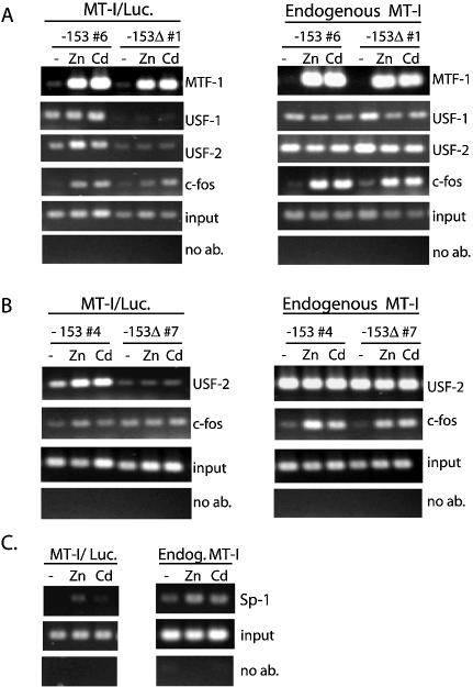
Figure 7.
Mapping interactions of transcription factors (MTF-1, USF, c-fos and Sp1) with MT-I promoter in vivo. ChIP analyses, using the stably- transfected Hepa cell lines –153-Luc-6 and –153Δ-Luc-1 (A), or –153-Luc-4 and –153Δ-Luc-7 (B), were carried out using the indicated antibodies and primers specific for the endogenous MT-I gene (right panels) or the integrated reporter genes (left panels). Cells remained untreated (–) or were treated for 2 h with zinc (100 µM) or cadmium (10 µM). For the number 4 and number 7 cell lines, ChIP results from USF-1 and MTF-1 immunoprecipitations (not shown) were similar to those shown for the number 6 and number 1 cell lines, respectively (A). In (C), chromatin was isolated from the –153-Luc-4 cells that were treated with zinc or cadmium for 2 h, ChIP carried out using an antibody against Sp1, and PCRs performed using primers to the transfected –153-Luciferase construct (MT-I/Luc) or the endogenous MT-I gene (Endog. MT-I).

