
Figure 1.
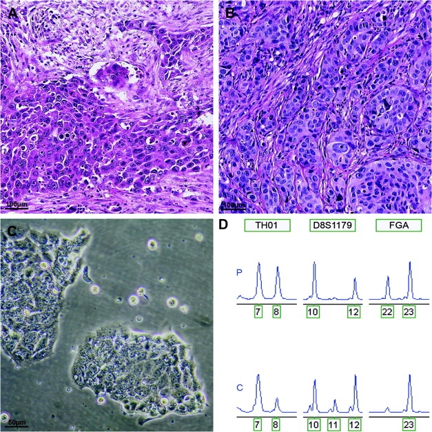
Authentication of human esophageal adenocarcinoma cell line OE33. A and B) Hematoxylin- and eosin-stained sections of the original tissue and xenograft of OE33 showing a poorly differentiated adenocarcinoma. C) In vitro growth pattern of cell line OE33. D) Short tandem repeat profile of the primary normal tissue (P) and cell line OE33 (C) indicating correct derivation of the cell line. Short-term repeat loci are indicated in boxes above electropherogram; the number of repeat units is indicated below the peaks. Note: loss of heterozygosity in the cell line at loci TH01 and FGA. The additional allele (11 repeat units) of D8S1179 observed in cell line OE33 is a known phenomenon and is probably due to somatic mutation or localized chromosomal rearrangements at this heterozygous locus.