
Figure 8:
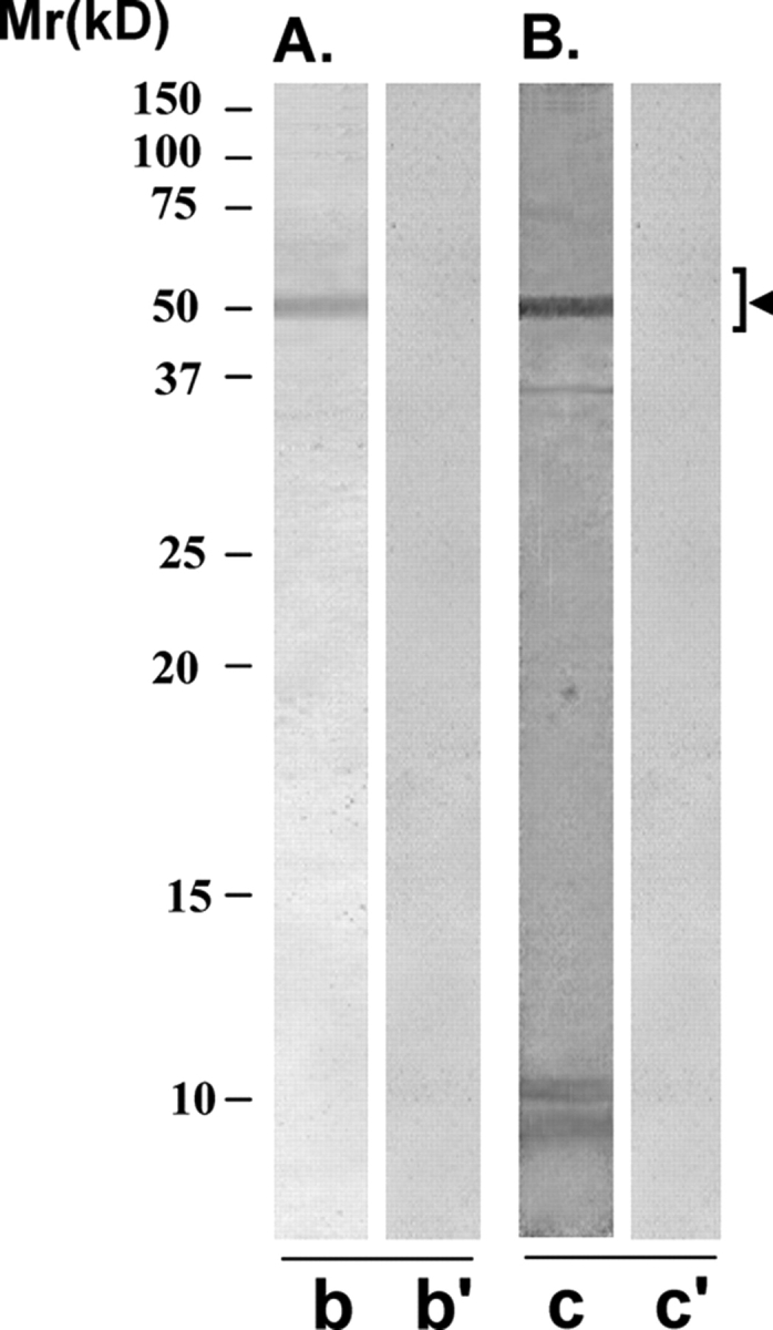
Immunoreactivity pattern of YLP20 scFv antibody with HSE
The purified scFv was examined for its reactivity with LIS-solubilized HSE using western blot (A) and immunoprecipitation (B) procedures. (A) YLP20 scFv antibody specifically recognized a protein band of 48 ± 5 kDa, corresponding to YLP12 antigen, on western blot of HSE (lane b). Control scFv antibody did not react with any specific band on the western blot (lane b’). (B) YLP20 scFv antibody Sepharose 4B immunobeads reacted with a specific protein in HSE that on elution with glycine–HCl (0.1 M, pH 2.8) showed a single band of 48 ± 5 kDa in SDS–PAGE (lane c). Control scFv antibody Sepharose 4B immunobeads did not react with any protein in HSE (lane c’).