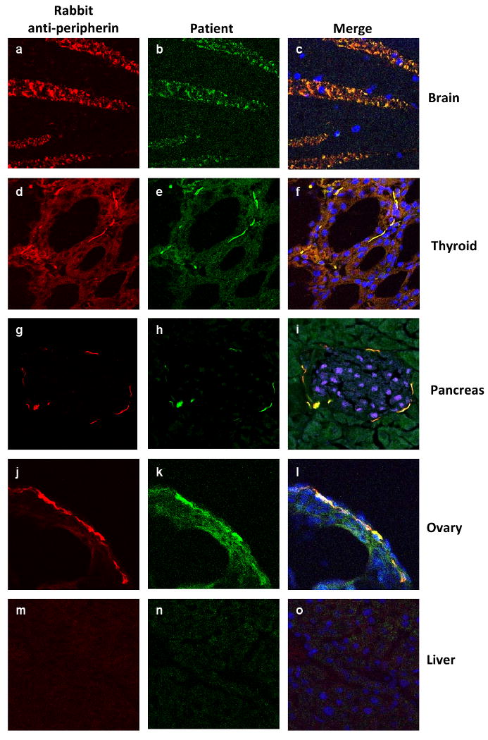
Fig. 5.

Patient IgG colocalizes with peripherin immunoreactivity in brain and endocrine organs. Tissues (brain [a-c], thyroid [d-f], pancreas [g-i], ovary [j-l] and liver [m-o]) were harvested from a 6-8 week old female mouse, cryosections (8 μm) were cut and stained with rabbit anti-peripherin-IgG (left columns), and patient IgG (center columns). All merged images (right column) show nuclear DAPI staining (blue) except for pancreas, where endocrine islet cells (i) were identified by IgG specific for the β-cell transcription factor PDX-1 (pseudo-colored purple).
