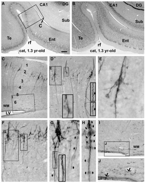
Fig. 2.
Doublecortin labeling in the entorhinal and adjoining temporal cortex of a young adult cat, focusing on clusters and chain-like formations. Panels A and B are low power views of immunolabeling and Nissl stain, respectively. Immunolabeled profiles in the framed area in (A) are enlarged through (C–H). The clusters are consisted of densely packed small cells. They reside at the border of layers I and II, and many of them form the bases of migratory cells (D, F). Many cells appear to migrate away from the base in a radiation manner (E, G), other cells and processes arrange as chains extending inwardly as far as layer V even VI. For a given chain, cells (small fat arrows) line up tightly in the proximal segment, but are separated by processes in the distal segment with increasing distance towards the end of the chain (F, G). A short arm extends from the base into layer I, which is consisted of immunoreactive processes but not somata (D–F). Panels I and J show a few labeled cells (arrows) in the ventricular wall and adjacent white matter (WM). Scale bar = 600 μmin A applying to B; equal to 200 μm for C; 100 μm for D, F, I; 50 μm for G, J and 25 μm for E, H.

