Fig. 3.
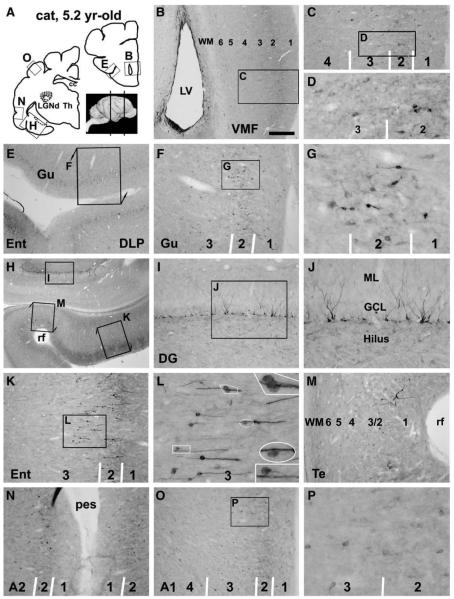
Doublecortin immunoreactive (DCX+) cells in representative forebrain areas in a 5.2 yr-old cat, as indicated in panel A. Panels B–G illustrate occurrence of some DCX+ cells in the ventral frontal areas, mostly in layers II/III. These cells are in bipolar or multipolar shape, range from small to medium size with distinct to weak reactivity (D, G). Note the reduced immunolabeling in the subventricular zone lining the anterior horn (B), relative to Fig. 1C. Panel H–M show labeled profiles in the dentate gyrus and the transitional area between the entorhinal and temporal cortices. Some DCX+ granule cells are present in the dentate gyrus with their cell bodies located at the subgranular zone (I, J). Small to medium-sized cells are present over layers II–III in the entorhinal cortex, mainly arranged as radially oriented chains. Some cells appear to leave the chain and migrate towards deeper locations (L, with enlarged areas as inserts). Both distinctly and lightly stained DCX+ cells remain in the low temporal (M) and auditory II (N) areas with low density. In more dorsally located auditory I area, only a small number of weakly labeled cells (medium-size) are present (O, P). Scale bar = 750 μm in B applying to E, equal to 1.5 mm for H, 300 μm for C, F, I, K, M–O; 100 μm for D, G, L, P and 33 μm for the enlarged inserts in L.

