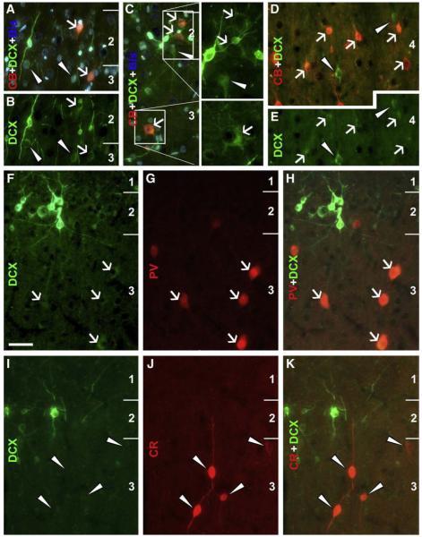
Fig. 6.
Colocalizations of calcium-binding proteins in DCX+ cells in the temporal neocortex of an adult cat (3.5 yr-old). Panels A–C show a partial colocalization (white arrows) of calbindin (CB) in medium (A, B) to large (C, with boxed areas enlarged 2× on the right) DCX cells in layers II/III. Many medium-sized DCX+ cells in deep layers are also co-labeled for CB (white arrows, D, E), but small and some large DCX+ cells lack CB reactivity (white arrowheads, A–E). Panels F–H show parvalbumin (PV) reactivity in weakly stained DCX+ cells in layer III (white arrows). Calretinin (CR) immunoreactive neurons do not exhibit DCX labeling (I–K). Scale bar = 50 μm in F, applying to other panels.

