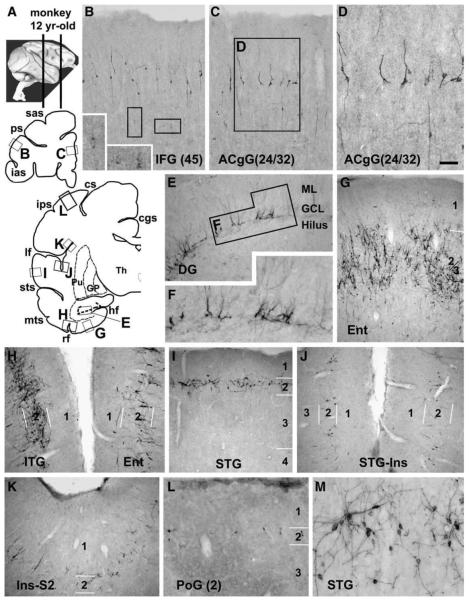
Fig. 8.
Doublecortin immunoreactive (DCX+) cells in representative adult monkey (Macaca mulatta) forebrain areas, as marked on the upper-left hemispheric maps (A). Panels B–D illustrate DCX+ cells in the prefrontal areas. Distinctly and weakly stained cells are present in layers II and upper III, with some weakly stained cells also reside in deeper locations. Panels E and F show moderate labeling in dentate granular cells. Panels G–L show a ventrodorsal gradient of labeling across the hemisphere over the temporoparietal cortex. Labeled cells in the entorhinal cortex arrange according to the island formations (G). Only a few labeled cells are detected in the dorsally-located somatosensory cortex (L). At higher magnification, these cells are in bipolar or multipolar shape, with varying somal size and labeling intensity (D, M). Scale bar = 150 μm in D applying to M, equal to 300 μm for B, C, E, G–L and 100 μm for M.

