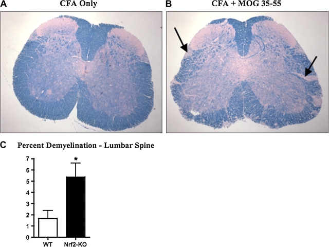
FIG. 2.
Nrf2-KO mice have greater demyelination than WT mice with EAE. (A and B) LFB staining of representative paraffin-embedded sections from lumbar spine of nondiseased (left panel) and diseased (MOG-induced EAE, right panel) mice. Bolded black arrows point to areas exemplifying demyelination. (C) Quantification of demyelination of sections described in (A and B) using ImageJ processing software and represented as mean ± SEM (WT, n = 4; Nrf2-KO, n = 5). *Significantly different than WT control (p < 0.05).

