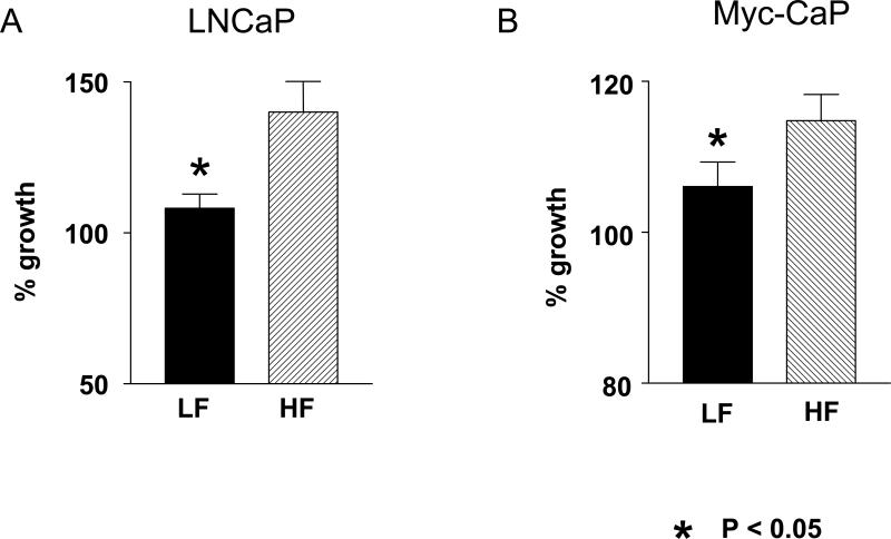
Figure 3. LNCaP and Myc-CaP proliferation in media containing HF and LF serum.
A. LNCaP cells had less proliferation in media containing the LF group mouse serum compared to the cells grown in media containing the HF group serum from Hi-Myc mice. The cell proliferation was measured after 48 hour incubation in the media containing 10 % mouse serum. N=8 for each diet group.
B. Myc-CaP cells grown in media containing 10 % Myc-mouse serum had less proliferation with LF group serum compared to the serum from HF diet group after 24 hour incubation. N= 5 for each group
All experiments were performed in triplicate with the serum from individual mice (not pooled). Data is expressed as a percentage of the cell growth in media containing 10 % FBS. Values are means ± SEM. *P<0.05. Proliferation was determined by measuring colorimetric changes of MTS tetrazolium compound into colored formazan product by NADPH or NADH produced by active cells.

