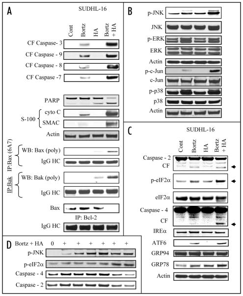
Figure 2.
Combined exposure to bortezomib and HA14-1 leads to a dramatic increase in caspase activation, mitochondrial damage, Bax and Bak translocation and conformational change, in association with JNK activation and ER stress induction in SUDHL16 cells. SUDHL16 cells were treated with 3 nM bortezomib ± 3.0 μM of HA14-1 for 14 h. (A) cytosolic (S-100) fractions were obtained as described in Materials and Methods, and expression of cytochrome c, AIF and Smac/DIABLO were monitored by western blot. Proteins from whole cell lysates were prepared and expression of the indicated proteins were determined by western blotting. Bax and Bak translocation and conformational change, as well as the association between Bax and Bcl-2 were monitored by immunoprecipitation followed by western blotting as described in Methods (B and C). At the end of the drug exposure (14 h) as (A) above, cells were lysed, sonicated, the proteins denatured, and subjected to western blot analysis using the indicated primary antibodies. (D) SUDHL 16 cells were treated with 3 nM bortezomib ± 3 μM HA14-1 for various intervals and changes in the expression of the indicated protein expression were monitored by western blotting. For these and all other studies, each lane was loaded with 30 μg of protein; blots were stripped and reprobed with antibodies directed against actin to ensure equivalent loading and transfer. Results are representative of three separate experiments.

