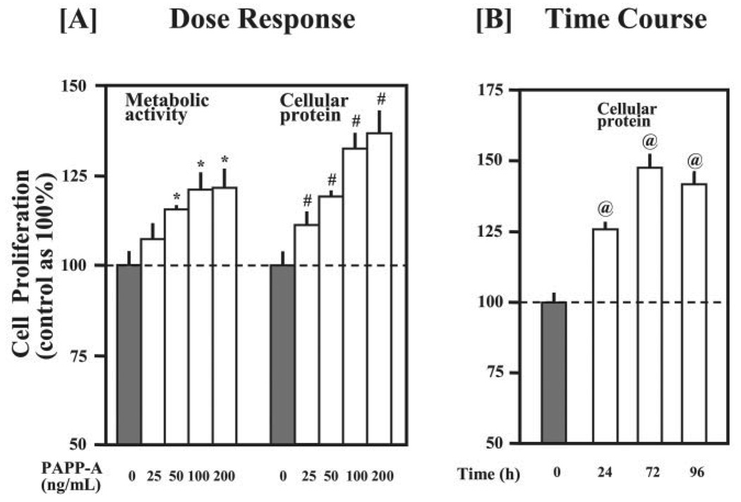
FIGURE 2. Effect of PAPP-A on the proliferation of C2C12 myoblasts.
Cellular proliferation was first determined by AlamarBlue assay, followed by quantitation of cellular proteins by Bradford reagent in the cell lysates as described under “Experimental Procedures.” A, C2C12 myoblasts were cultured for 48 h in DMEM containing 5% FCS and varying amounts of purified recombinant PAPP-A protein. The data presented here show that PAPP-A treatment increases the proliferation of C2C12 myoblasts in a dose-dependent manner (*, p<0.05 versus untreated control and #, p<0.05 versus untreated control). B, C2C12 myoblasts were seeded in 24-well plates (10,000 cells/well) and treated with PAPP-A (100 ng/ml). After PAPP-A treatment, cellular proliferation was measured at the indicated time points. The data show that the maximum proliferation of C2C12 myoblasts was noticed after 72 h of PAPP-A treatment (@, p <0.05 versus 0 h).

