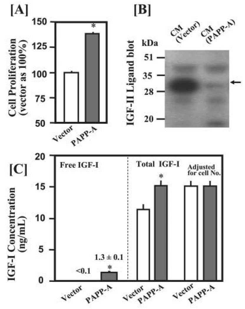
FIGURE 7. PAPP-A overexpression increases free IGF-I concentration primarily through enhancing degradation of the IGFBP-2 produced by C2C12 myoblasts.
C2C12 cells were seeded in 5% CS in 6-well plates at a density of 80,000 cells/well. After 24 h of incubation, cells were transfected with 1 µg of pFLAG empty vector DNA or PAPP-A-(1–1547)/pFLAG plasmids in 1 ml of DMEM containing 5% CS. After 24 h of incubation, medium was replaced with 2 ml of DMEM containing 5% CS. CM samples were collected after an additional 48 h of incubation for determination of IGFBP proteolysis and IGF-I concentrations. A, overexpression of PAPP-A significantly increased cell proliferation as determined by the AlamarBlue assay. B, 40 µl of CM were subjected to IGF-II ligand blot analysis. Overexpression of PAPP-A caused a nearly complete degradation of the mIGFBP-2 (indicated by arrow) produced by C2C12 myoblasts. C, 50 µl of CM was used to determine free IGF-I concentrations by a commercial kit (Diagnostic Systems Laboratories, Webster, Texas). Total IGF-I concentrations were determined by radioimmunoassay (29). Similar results were obtained in another independent experiment. *, p <0.05 versus vector control (n = 6)

