
Figure 1.
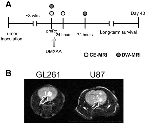
(A) Schematic outline of the study design. Approximately 3 weeks post intracranial implantation of GL261 and U87 cells, tumor-bearing mice were administered a single dose of the VDA DMXAA as described in the Materials and Methods section. CE-MRI was performed prior to (preRx) and 24 hours following treatment to monitor the vascular response of gliomas to DMXAA treatment. Additionally, DW-MRI was performed 72 hours post treatment to examine changes in cellularity following VDA therapy. Long-term survival analysis was performed by observing animals in the control and treatment groups over a 40-day period. (B) T2W MRI of tumor growth. T2-weighted (T2W) MR images of a control C57Bl6 mouse bearing a GL261 glioma and a nude mouse bearing U87 glioma are shown. Tumors appeared as well-defined hyperintense regions at the site of injection as indicated by the arrows.